Nuclear Medicine is a specialized form of medical imaging and treatment that utilizes the energy released from the nucleus of an atom. Nuclear Medicine provides targeted imaging and therapy through the unique distribution properties of radioactive materials used in both diagnostic and therapeutic applications.
For imaging, a very small amount of radioactive material is administered, which travels to specific organs or tissues in the body. The radiation emitted is detected and used to create detailed images that "highlight" the targeted lesion, distinguishing it from surrounding healthy tissues. This allows for highly precise imaging of areas that may be affected by disease or dysfunction, even at an early stage, when other methods might not yet detect any structural changes.
In therapy, the radioactive material is directed specifically to the targeted lesion, minimizing the effect on surrounding healthy tissues. This targeted approach allows nuclear medicine to treat various conditions, especially cancers, with great precision.
Why Choose Nuclear Medicine?
Early Detection of Functional Changes
Unlike traditional imaging methods, nuclear medicine can detect functional changes in tissues and organs before any anatomical abnormalities become apparent. This enables earlier diagnosis of conditions, improving treatment outcomes.
Comprehensive Imaging
When combined with conventional imaging techniques such as CT and MRI, nuclear medicine provides a comprehensive view of both the anatomical and functional aspects of a patient's health. This holistic approach ensures the most accurate and detailed information is available for effective diagnosis and treatment.
Safe and Effective
Nuclear medicine imaging is generally safe and involves a minimal amount of radiation. The information gained from these procedures can offer invaluable insights into a patient's health, leading to better-informed treatment decisions.
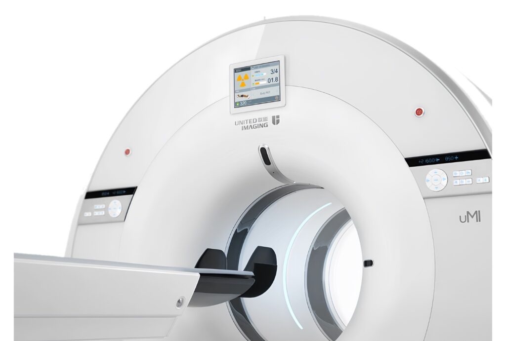
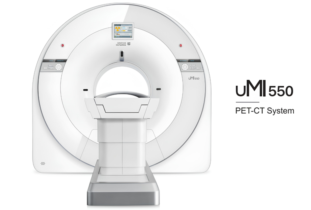
Positron Emission Tomography (PET) - PET/CT SCAN
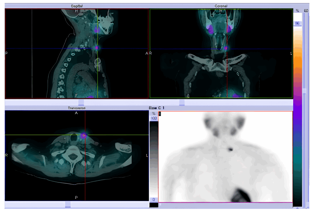
Positron Emission Tomography (PET) combined with Computed Tomography (CT) provides a unique, non-invasive, and highly accurate imaging technique that allows clinicians to observe functional changes in the body. This advanced imaging method offers both detailed anatomical and metabolic information, giving a comprehensive picture of a patient's health.
PET works by using a small amount of radioactive material to track metabolic activity within the body. The most commonly used radioactive tracer worldwide is 18F-FDG, a glucose analog. This substance is preferentially absorbed by cells with higher metabolic activity, such as cancer cells. Therefore, areas with abnormal or increased metabolic activity, such as tumors, will appear "hotter" (highlighted) in the scan.
The procedure itself is safe, with relatively low radiation exposure, and can provide invaluable information for diagnosing and monitoring various conditions.
Indications for PET/CT Scan
PET/CT scans are used for both oncological and non-oncological purposes, making them a versatile tool for comprehensive healthcare management.
Oncological Indications:
Cancer Management: PET/CT scans are beneficial in the staging of cancer, assessing the extent of the disease, and evaluating how well a tumor is responding to treatment. It is also crucial for restaging and surveillance to monitor for potential recurrence.
Non-Oncological Indications:
Dementia: PET/CT can help assess brain function, aiding in the diagnosis of conditions like Alzheimer’s disease and other forms of dementia.
Epilepsy: For patients with epilepsy, PET/CT can help pinpoint the origin of seizures and guide appropriate treatment options.
Fever of Unknown Origin: In cases where the cause of fever cannot be determined, PET/CT imaging can help identify underlying infections or inflammatory diseases.
Cardiac Viability: PET/CT scans are used in cardiology to evaluate heart function, particularly in assessing areas of the heart that may still be viable after a heart attack.
This combination of functional and structural imaging makes PET/CT an essential tool in diagnosis, treatment planning, and follow-up care for a wide range of medical conditions.
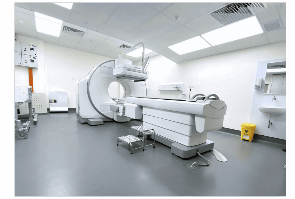
Single Photon Emission Computed Tomography (SPECT-CT)
Single Photon Emission Computed Tomography (SPECT-CT) is a hybrid imaging technique that combines the functional imaging capabilities of SPECT with the detailed anatomical information from CT. This advanced imaging modality provides sensitive, precise, and targeted insights into how different parts of the body are functioning.
While similar to PET-CT, SPECT-CT has the advantage of being able to perform a broader range of imaging studies, using different types of radioactive tracers to assess various physiological processes. Like PET-CT, the procedure is very safe, and the radiation exposure from the scan is relatively low.
Types of SPECT-CT Scans
SPECT-CT offers a variety of imaging applications, particularly in cardiology, orthopedics, nephrology, and oncology, among others. Some of the most common SPECT-CT scans include:
Heart Imaging:
Perfusion Studies: Evaluates blood flow to the heart muscle, helping detect blockages or areas at risk of damage.
Viability Studies: Assesses the health of heart tissue, particularly after a heart attack.
Ejection Fraction Studies: Measures the heart’s ability to pump blood and assess overall heart function.
Bone Imaging: Identifies areas of bone inflammation, infection, or abnormalities, such as in cases of fractures, infections, or bone cancer.
Renal Imaging:
DTPA/MAG3/DMSA Scans: Used to assess kidney function, detect obstructions, and evaluate overall kidney health and perfusion.
Lung Perfusion Imaging: Evaluates blood flow in the lungs, helping detect conditions like pulmonary embolism or other pulmonary disorders.
Gastrointestinal-Related Imaging:
Meckel’s Scan: Identifies abnormalities in the intestines, such as Meckel’s diverticulum.
HIDA Scan: Assesses gallbladder function and helps diagnose conditions like cholecystitis.
Bleeding Detection: Detects active gastrointestinal bleeding sources.
Endocrine Imaging:
Parathyroid Imaging: Helps locate abnormal parathyroid glands, especially in the case of hyperparathyroidism.
Thyroid I-131 Scan: Used to evaluate thyroid function, particularly after thyroid cancer treatment.
Whole Body I-131 Post-Therapy Imaging: Used for post-therapy monitoring of thyroid cancer, assessing the spread of cancer cells or the effectiveness of treatment.
This hybrid imaging technique allows for a comprehensive and targeted approach to diagnosing and monitoring a wide variety of conditions, enhancing clinical decision-making and patient care.
Why Choose SPECT-CT?
Versatility
SPECT-CT can assess a wide range of medical conditions, from heart disease to bone disorders, providing crucial information that other imaging methods may not reveal.
Precision
Combining functional imaging with detailed anatomical scans, SPECT-CT provides precise insights into the location and extent of diseases, aiding in accurate diagnosis and treatment planning.
Safety
The procedure is non-invasive, safe, and associated with low radiation exposure, making it an ideal option for frequent monitoring and diagnostics.
Radioiodine-131 Therapy
A type of nuclear medicine treatment that uses radioactive iodine to target and destroy thyroid cells. This therapy is primarily used to treat hyperthyroidism and thyroid cancer.
Treatment for Hyperthyroidism
Hyperthyroidism occurs when the thyroid gland produces an excess of hormones, either due to hyperfunctioning nodules or overactive thyroid tissue. Radioiodine-131 is uniquely effective because it targets and destroys these hyperfunctioning thyroid cells while sparing the healthy thyroid tissue. As a result, this therapy helps restore normal thyroid function by reducing hormone production to appropriate levels.
Treatment for Thyroid Cancer
In the case of thyroid cancer, Radioiodine-131 therapy is commonly used after the surgical removal of the thyroid gland. The therapy works by targeting and eliminating any remaining thyroid cells or metastatic thyroid tissue that might have spread to other parts of the body. This minimizes the risk of cancer recurrence and helps ensure that any residual cancerous cells are destroyed.
How it Works
The radioactive iodine is typically administered orally in the form of a capsule or liquid. It is absorbed by the body and specifically targeted to the thyroid tissue. Once the iodine reaches the thyroid, it emits radiation that destroys the targeted thyroid cells. Since it is a targeted therapy, only the thyroid tissue is affected, and surrounding healthy tissues are spared.
Safety and Effectiveness
Radioiodine-131 therapy is highly effective and is safely administered under the supervision of a Nuclear Medicine Physician. The procedure is usually completed in a single dose for each treatment cycle. Because it is a targeted therapy, the side effects are minimal, and the risk of secondary cancers is very low.
Benefits of Radioiodine-131 Therapy
Minimal Side Effects: As a targeted therapy, it primarily affects the thyroid tissue, sparing surrounding healthy tissues and minimizing side effects.
Effectiveness: Radioiodine-131 is a proven and effective treatment for both hyperthyroidism and thyroid cancer, with high success rates in preventing recurrence.
Non-invasive: The treatment is non-invasive and requires only a single oral dose for each treatment cycle.
Radioiodine-131 therapy is an essential part of managing thyroid disorders, offering a safe, effective, and minimally invasive solution. If you have any concerns or questions about the therapy, your Nuclear Medicine physician will guide you through the process and provide personalized care
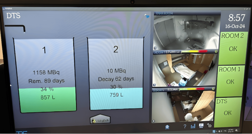
Isolation Ward Room
The Isolation Ward Room for High Dose Radio-Iodine Therapy is well equipped with shielding, advanced remote radiation monitoring system and hot toilet that connects to shielded decay tank.
Advanced Systems & Facilities
Remote Monitoring System
Real-time monitoring system that tracks radiation levels and room status remotely.
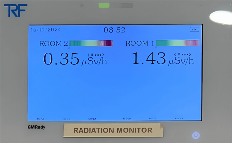
Radiation Monitor
Advanced monitors provide accurate, real-time radiation data for a safe environment
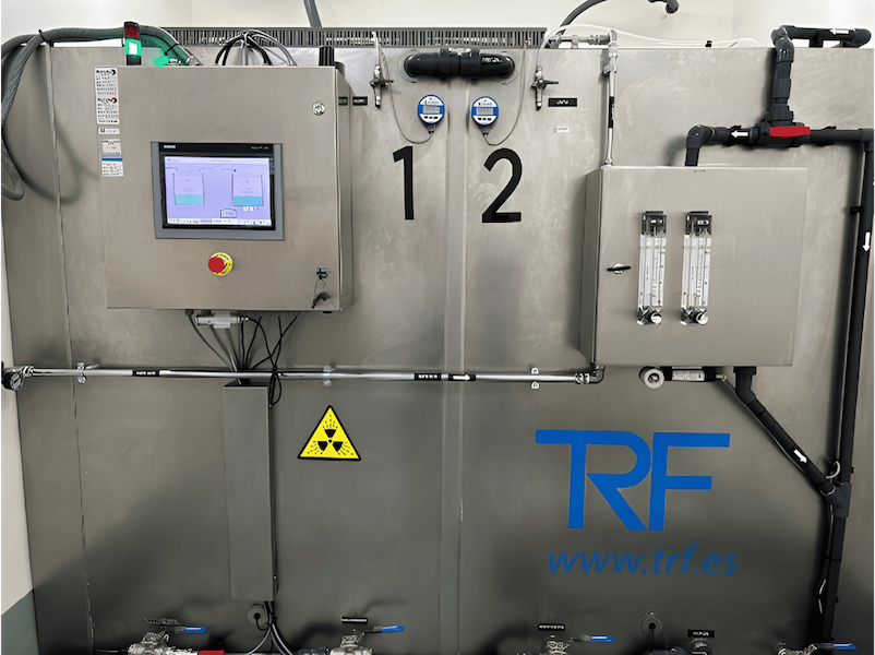
Shielded Decay Tank System
Safely transfer and stores radioactive in a shielded decay tank
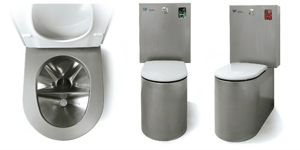
Hot Toilet with Decay Tank
Specially designed hot toilet connected to the decay tank system for safety and hygiene
Prostate Cancer Therapy – Lutetium-177 PSMA
Lutetium-177 PSMA (Prostate-Specific Membrane Antigen) Therapy is an advanced form of nuclear medicine therapy designed to target and treat metastatic prostate cancer. This therapy uses Lutetium-177, a radioactive isotope, to specifically target prostate cancer cells that have spread to other areas of the body, such as lymph nodes and bones.
The treatment works by attaching the radioactive Lutetium-177 to a molecule that binds to PSMA, a protein found in high amounts on the surface of prostate cancer cells. Once the Lutetium-177 is delivered to the cancer cells, it emits radiation that destroys these cancerous cells while minimizing damage to surrounding healthy tissues.
How Lutetium-177 PSMA Therapy Works
Lutetium-177 PSMA therapy is targeted and precision-based, meaning that it focuses specifically on the metastatic prostate cancer cells, making it an effective treatment for patients with advanced prostate cancer. It is especially useful for cases where cancer has spread to other parts of the body, such as the bones or lymph nodes.
Benefits of Lutetium-177 PSMA Therapy
Effective Treatment for Metastatic Prostate Cancer: Lutetium-177 PSMA therapy is highly effective for patients with prostate cancer that has spread to other parts of the body, especially the bones and lymph nodes.
Prolongs Life Expectancy: The therapy has been shown to prolong survival in patients with advanced prostate cancer, providing a promising treatment option for those with limited options.
Alleviates Symptoms: By targeting and destroying metastatic prostate cancer cells, this therapy can significantly reduce symptoms related to metastasis, including bone pain and discomfort.
Minimally Invasive: Lutetium-177 PSMA therapy is a non-invasive procedure that involves intravenous administration of the radioactive substance, offering a less invasive alternative to traditional treatments such as surgery or radiation.
Precision Medicine: The therapy is highly targeted, meaning that the radioactive material is delivered directly to the cancerous cells, minimizing the risk of damage to surrounding healthy tissues.
Safety and Supervision
Lutetium-177 PSMA therapy is administered under the close supervision of an experienced Nuclear Medicine Physician. The procedure is safe, and radiation exposure is controlled to minimize risks. It is a promising treatment option for patients with advanced prostate cancer who have not responded well to other therapies.
This innovative treatment offers hope and improvement in quality of life for patients battling metastatic prostate cancer, by focusing on targeted therapy with minimal side effects.
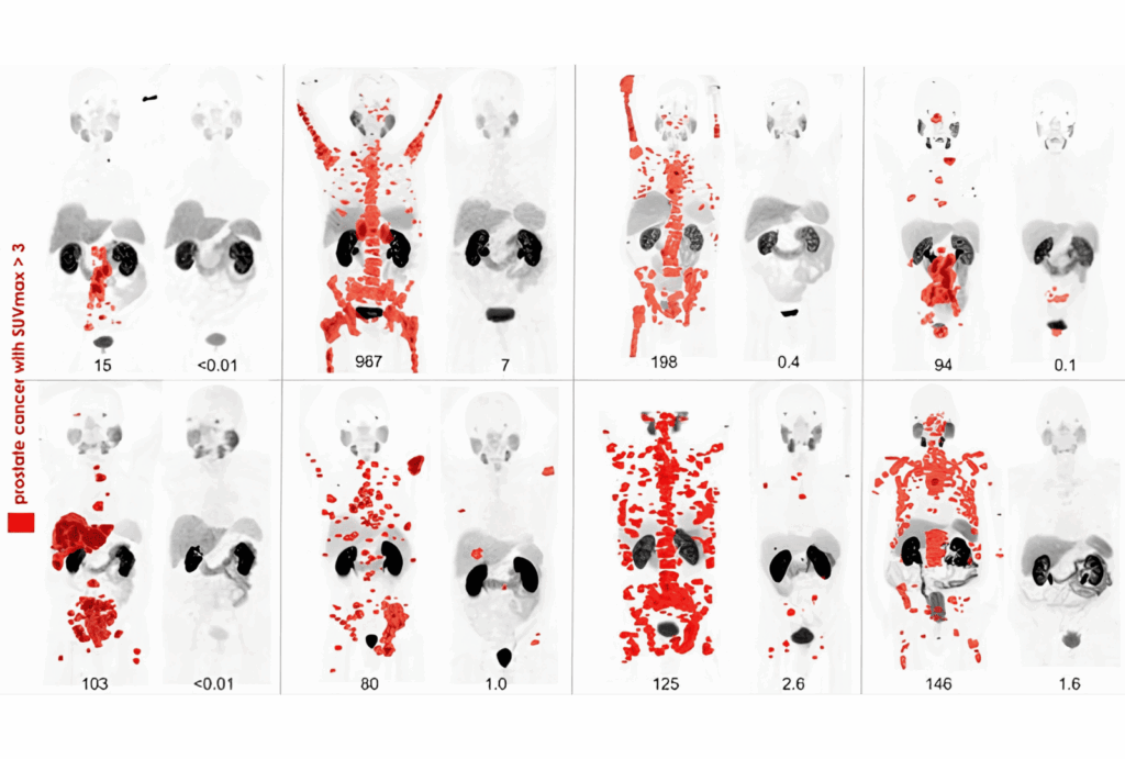
2018 Annual Meeting of the Society of Nuclear Medicine and Molecular Imaging (SNMMI) Image of the Year Award,
PSMA PET before and after lutetium-177 PSMA617 theranostics in 8 patients with metastatic prostate cancer who exhausted standard therapeutic options.
68Ga-PSMA11 PET maximum intensity projection (MIP) images at baseline and 3 months after 177Lu-PSMA617 in 8 patients with PSA decline ≥ 98 percent in a prospective phase II study. Any disease with SUV over 3 is in red. Credit: Michael Hofman, John Violet, Shahneen Sandhu, Justin Ferdinandus, Amir Iravani, Grace Kong, Aravind Ravi Kumar, Tim Akhurst, Sue Ping Thang, Price Jackson, Mark Scalzo, Scott Williams and Rodney Hicks, Peter MacCallum Cancer Centre, Melbourne, Australia.
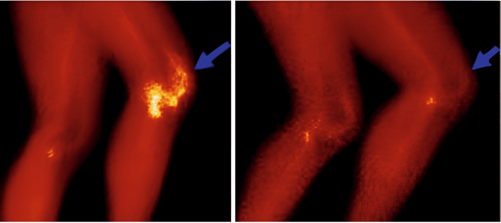
Radiosynovectomy for Joint Disorders
Radiosynovectomy is a targeted nuclear medicine treatment used to treat painful joints caused by synovitis—inflammation of the synovial membrane, which lines the joints. This therapy involves the injection of a radioactive substance directly into the affected joint. The radioactive material is carefully selected to ensure it only travels a very short distance which means it can effectively destroy the inflamed synovial tissue without causing damage to surrounding healthy tissues.
The treatment works by targeting the abnormal synovial cells, destroying the inflamed tissue, and thus reducing joint inflammation, pain, and deformity. Radiosynovectomy can significantly improve mobility and quality of life for patients suffering from chronic joint pain caused by diseases like rheumatoid arthritis or other forms of inflammatory arthritis.
Benefits of Radiosynovectomy
Targeted Treatment: The radioactive material is injected directly into the joint, where it specifically destroys the inflamed tissue, minimizing damage to surrounding healthy structures.
Minimal Side Effects: Because the radioactive material only travels a short distance, there is minimal exposure to surrounding healthy tissues, reducing the risk of side effects.
Long-Term Relief: Radiosynovectomy can provide lasting relief from joint pain, reduce swelling, and help preserve joint function, allowing patients to regain mobility.
Intraarticular injection of radionuclides, called radiosynovectomy, radiosynoviorthesis is performed to treat the synovial inflammation alternatively to surgery.
Bone Palliation Radionuclide Therapy
Bone Palliation Radionuclide Therapy is used to treat bone metastases—a common complication of cancers such as prostate cancer, breast cancer, and other solid tumors. Bone metastasis can be particularly painful, affecting the bones and causing significant discomfort for patients. When metastasis is widespread, treatment options are focused on relieving pain and improving quality of life.
In cases of extensive bone metastases, radiopharmaceuticals such as strontium-89, radium-223, and samarium-153 are used for targeted therapy. These radioactive substances are selectively absorbed by bone tissue, where they emit radiation that targets and destroys the cancerous cells, helping to alleviate bone pain associated with metastasis. This form of therapy is effective in reducing symptoms, improving mobility, and enhancing overall quality of life for patients.
Types of Radiopharmaceuticals Used:
Strontium-89: Targets areas of bone metastasis, particularly in the pelvis, spine, and long bones, providing pain relief and slowing cancer progression.
Radium-223: Primarily used for prostate cancer, this radiopharmaceutical selectively targets bone metastases and delivers localized radiation to destroy cancer cells while minimizing damage to surrounding healthy tissue.
Samarium-153: Similar to strontium-89, samarium-153 helps relieve bone pain and is commonly used for patients with widespread bone metastasis.
Benefits of Bone Palliation Radionuclide Therapy:
Effective Pain Relief: The therapy is highly effective in providing relief from bone pain caused by metastases, allowing patients to manage discomfort and improve their quality of life.
Targeted Treatment: The radiopharmaceuticals are specifically absorbed by the bone, concentrating the radiation on cancerous areas, which minimizes exposure to healthy tissues.
Minimal Side Effects: Bone palliation therapy has a relatively low risk of side effects compared to other more invasive treatments like surgery or radiation therapy.
Safety and Supervision
Both Radiosynovectomy and Bone Palliation Radionuclide Therapy are safe, non-invasive treatments administered under the supervision of a Nuclear Medicine Physician. These therapies are designed to improve the quality of life for patients, providing targeted relief with minimal risks and side effect
Contact info
Please make appointment 2 weeks in advance to avoid disappointment.